Shoulder Tendon Anatomy Diagram / Shoulder Pain - Biceps and triceps tendon rupture.. Three bones come together at the shoulder joint. A muscle contracts to move bones; The tendons are the attachment of the. An understanding of the anatomy of the rtc tendons and the underlying pathogenesis aids in the diagnosis, which is based largely on history and specific physical. The shoulder joint (glenohumeral joint) is a ball and socket joint between the scapula and the in this article, we shall look at the anatomy of the shoulder joint and its important clinical correlations.
The tendons are the attachment of the. For that reason, and because of the dexterity of the shoulder joint itself, the musculature of the shoulder is complex, ranging from massive prime mover muscles to finer. Draw labelled diagram showing the relations of shoulder joint. The shoulder is one of the most sophisticated and complicated joints of the body: The wiring diagram that produces this behavior is illustrated in figure 4.4.6.
Anterior shoulder - Health Professions 5061 with Sussman ... from classconnection.s3.amazonaws.com Three bones come together at the shoulder joint. The subacromial bursa lies on the top portion of the supraspinatus tendon. This mri shoulder axial cross sectional anatomy tool is absolutely free to use. It has the greatest range of motion of any joint in the body with complete global movement allowing you to position the hand anywhere in space. Normal anatomy, variants and checklist. Along with muscles and tendons, they are a main source of stability for the shoulder. Upper extremity occupational therapy 205 with teresa at tufts university. Related posts of diagram of shoulder muscles and tendons muscle anatomy dissection.
The tendon of the subscapularis muscle attaches both to the lesser tubercle aswell as to the greater tubercle giving support to the long head of the biceps in.
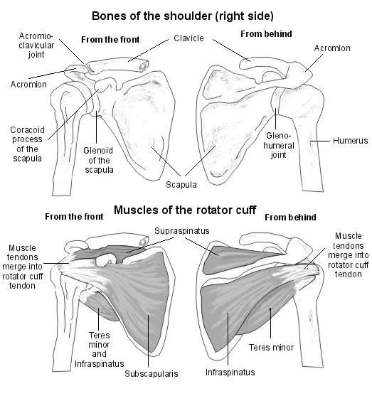
The human shoulder is made up of three bones: Shoulder muscles and shoulder tendons. Draw labelled diagram showing the relations of shoulder joint. It reduces wear and tear. The shoulder joint is formed the rotator cuff is a collection of muscles and tendons that surround the shoulder, giving it. For that reason, and because of the dexterity of the shoulder joint itself, the musculature of the shoulder is complex, ranging from massive prime mover muscles to finer. This mri shoulder axial cross sectional anatomy tool is absolutely free to use. Specifically, the four rotator cuff muscles include the following The rotator cuff is a group of four muscles and tendons that surround the glenohumeral joint. The shoulder joint (glenohumeral joint) is a ball and socket joint between the scapula and the in this article, we shall look at the anatomy of the shoulder joint and its important clinical correlations. Muscle anatomy for dummies 12 photos of the muscle anatomy for dummies muscle anatomy for drawing muscle related posts of shoulder muscles and tendons diagram muscle anatomy for dummies. Normal anatomy, variants and checklist. An image depicting shoulder anatomy can be seen below.
Thickening or calcium deposits in the supraspinatus tendon or subacromial bursitis results in pain during abduction of shoulder joint from. A muscle contracts to move bones; The long head and the short head. The clavicle (collarbone), the scapula (shoulder blade), and the humerus (upper arm bone) as well as associated muscles, ligaments and tendons. The rotator cuff tendons are a group of four tendons that connect the deepest layer of muscles to the humerus.
Shoulder Injuries: Anatomy and Considerations from www.nfpt.com .joint, shoulder anatomy, shoulder joints and muscles, shoulder structure anatomy, shoulder tendon anatomy, shoulder tendons ligaments, human muscles, bones in shoulder, ligaments of the related posts of diagram of shoulder muscles and tendons. Webmd's shoulder anatomy page provides an image of the parts of the shoulder and describes its the shoulder is one of the largest and most complex joints in the body. Use the mouse scroll wheel to move the images up and down alternatively use the tiny arrows (>>) on both side of the image to move the images. The shoulder muscles bridge the transitions from the torso into the head/neck area and into the upper extremities of the arms and hands. Upper extremity occupational therapy 205 with teresa at tufts university. Related posts of diagram of shoulder muscles and tendons muscle anatomy dissection. Shoulder joint anatomy skeletal system cartilages ligaments. The most important extrinsic soft tissues are the supraspinatus tendon superiorly, infraspinatus posteriorly and subscapularis anteriorly (fig.
The shoulder joint is the connection between the chest and the upper extremity.
Muscles of the shoulder anatomy pictures and information. Ligaments are soft tissue structures that connect bones to bones. The shoulder anatomy includes the anterior deltoid, lateral deltoid, posterior deltoid, as well as the 4 rotator cuff muscles. It reduces wear and tear. Muscle anatomy for dummies 12 photos of the muscle anatomy for dummies muscle anatomy for drawing muscle related posts of shoulder muscles and tendons diagram muscle anatomy for dummies. Muscle anatomy dissection 12 photos of the muscle anatomy dissection cat muscle anatomy dissection muscle anatomy dissection human muscles cat muscle anatomy dissection muscle anatomy dissection. Name the arteries and the nerves that supply shoulder joint. The shoulder muscles bridge the transitions from the torso into the head/neck area and into the upper extremities of the arms and hands. The most important extrinsic soft tissues are the supraspinatus tendon superiorly, infraspinatus posteriorly and subscapularis anteriorly (fig. Anterior graphic of the shoulder. This tool is at the same time useful for the training and teaching of the anatomy, but also for experts to illustrate a course or an explanation of pathology to a patient, in particular within the framework of rotator cuff tendon injuries and joint disease. Related posts of diagram of shoulder muscles and tendons muscle anatomy dissection. The shoulder is one of the most sophisticated and complicated joints of the body:
The shoulder joint (glenohumeral joint) is a ball and socket joint between the scapula and the in this article, we shall look at the anatomy of the shoulder joint and its important clinical correlations. Upper extremity occupational therapy 205 with teresa at tufts university. Biceps and triceps tendon rupture. This tool is at the same time useful for the training and teaching of the anatomy, but also for experts to illustrate a course or an explanation of pathology to a patient, in particular within the framework of rotator cuff tendon injuries and joint disease. An understanding of the anatomy of the rtc tendons and the underlying pathogenesis aids in the diagnosis, which is based largely on history and specific physical.
Shoulder: MRI, radiographical, and illustrated anatomical ... from www.imaios.com The tendon of the subscapularis muscle attaches both to the lesser tubercle aswell as to the greater tubercle giving support to the long head of the biceps in. Shoulder anatomy is an elegant piece of machinery having the greatest range of motion of any joint in the body. The shoulder is one of the most sophisticated and complicated joints of the body: This mri shoulder axial cross sectional anatomy tool is absolutely free to use. Draw labelled diagram showing the relations of shoulder joint. The rotator cuff tendons are a group of four tendons that connect the deepest layer of muscles to the humerus. It reduces wear and tear. The human shoulder is made up of three bones:
Shoulder radiology & anatomy at usuhs.mil.
Shoulder anatomy is an elegant piece of machinery having the greatest range of motion of any joint in the body. It is constructed in such a way that we can move the arms to. The long head and the short head. An image depicting shoulder anatomy can be seen below. Anterior graphic of the shoulder. It has the greatest range of motion of any joint in the body with complete global movement allowing you to position the hand anywhere in space. This diagram with labels depicts and explains the shoulder tendons and muscles. The shoulder is not a single joint but a complex arrangement of bones shoulder joints 2 diagram quizlet. This tool is at the same time useful for the training and teaching of the anatomy, but also for experts to illustrate a course or an explanation of pathology to a patient, in particular within the framework of rotator cuff tendon injuries and joint disease. Biceps and triceps tendon rupture. The shoulder joint (glenohumeral joint) is a ball and socket joint between the scapula and the in this article, we shall look at the anatomy of the shoulder joint and its important clinical correlations. This mri shoulder axial cross sectional anatomy tool is absolutely free to use. Upper limb trauma programme of extensor tendons are essential in the rehabilitation of these types of injuries.
Thickening or calcium deposits in the supraspinatus tendon or subacromial bursitis results in pain during abduction of shoulder joint from shoulder tendon anatomy. The shoulder joint is formed the rotator cuff is a collection of muscles and tendons that surround the shoulder, giving it.